Medical Image Analysis with AI: Diagnostic Applications, Challenges, and Implementation Guide
Meta Description: Implement AI-powered medical image analysis for diagnostics. Learn applications, regulatory requirements, privacy concerns, and implementation best practices for healthcare.
Introduction: The Medical Imaging Revolution
Medical imaging produces approximately 30% of all healthcare data. X-rays, CT scans, MRI, ultrasound, and pathology slides contain critical diagnostic information. AI has transformed how we analyze these images, enabling faster diagnosis, detecting subtle patterns humans miss, and democratizing access to expert-level diagnostics.
In 2026, AI-assisted medical imaging is mainstream in many hospitals. Yet challenges remain: regulatory compliance, ensuring safety, managing liability, handling privacy-sensitive data, and maintaining performance on diverse patient populations. This comprehensive guide covers everything from technical implementation to regulatory navigation.
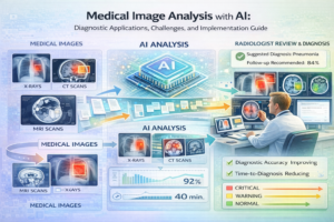
Medical Image Analysis Applications
1. Radiology: Chest X-Ray Analysis
One of the most widespread AI applications in medicine. AI systems detect pneumonia, tuberculosis, COVID-19, and lung cancer.
- Typical Accuracy: 92-96% sensitivity for pneumonia detection
- Clinical Impact: Reduces interpretation time from 5 minutes to 30 seconds; catches 15-20% more cases in screenings
- Implementation Cost: $50,000-$300,000 depending on integration complexity
- ROI Timeline: 12-18 months through efficiency gains and reduced liability
- Regulatory Status: Multiple FDA-approved solutions available (Zebra Medical, Subtle Medical)
Key Challenge: Handling diverse scanner manufacturers and acquisition parameters. Models trained on one hospital’s equipment may not generalize to another.
Solution: Transfer learning from large publicly-available datasets (CheXpert, MIMIC-CXR). Fine-tune on local hospital data (500-1,000 images).
2. Pathology: Cancer Diagnosis and Grading
Analyzing biopsy slides to detect cancer and determine severity. Some of the most critical AI applications in healthcare.
- Task: Detect cancer regions, classify tumor type, grade aggressiveness (Gleason score for prostate cancer)
- Accuracy: 95-98% when trained on adequate data
- Special Challenge: Whole Slide Images (WSI) are enormous (40,000×50,000 pixels). Can’t fit in GPU memory.
- Solution: Tile-based processing or attention-based multiple instance learning (MIL)
- Timeline to Deploy: 6-12 months for regulatory approval (FDA 510(k) typically required)
3. Ophthalmology: Diabetic Retinopathy Detection
Detecting disease in retinal fundus images. One of the earliest successful clinical AI deployments.
- Accuracy: 95%+ sensitivity, 98%+ specificity with adequate training data
- Clinical Impact: Screens can be automated, reducing burden on ophthalmologists by 80%
- Success Examples: Google’s screening programs in India and Southeast Asia (screened 2M+ patients)
- Key Success Factor: 128,000 annotated fundus images for training
- Deployment Model: Integrated into screening workflows, not replacing specialist review
4. Cardiology: Cardiac MRI Analysis
Automatically segmenting heart chambers and myocardium to assess function and detect abnormalities.
- Clinical Value: Reduces analysis time from 30 minutes to 2 minutes per patient
- Accuracy: Dice coefficient 92-96% (measure of segmentation accuracy)
- Implementation: U-Net style architectures with multi-modality fusion
- Challenge: Huge inter-patient variability in heart anatomy and size
5. Oncology: Tumor Detection and Monitoring
CT/MRI analysis for tumor detection, sizing, and response to treatment monitoring.
- Application: Detect new tumors, measure size changes (standard in clinical trials)
- Accuracy: 88-94% detection, but size measurement error critical (<5% required)
- Regulatory Requirement: FDA approval needed (typically 510(k) pathway)
- Current Status: Several FDA-approved solutions (Icad, IBM Watson for Oncology)
Technical Architecture for Medical Image AI
Typical Pipeline:
- Image Acquisition: Patient scan (DICOM format)
- Pre-processing: Anonymization, normalization, windowing (for CT/X-ray)
- AI Analysis: Deep learning model inference
- Post-processing: Refinement, confidence calibration
- Visualization: Highlight findings, uncertainty visualization
- Reporting: Generate clinical report with AI findings
Model Architectures for Medical Imaging
| Architecture | Task Type | Typical Accuracy | Advantages | Disadvantages |
|---|---|---|---|---|
| U-Net | Segmentation | 92-96% Dice | Excellent for small datasets, encoder-decoder symmetric | Limited context from limited receptive field |
| ResNet-50/101 | Classification | 92-96% accuracy | Proven, good with transfer learning, fast inference | Less sophisticated than modern vision transformers |
| Vision Transformer (ViT) | Classification/Detection | 93-97% accuracy | State-of-the-art accuracy, better at global patterns | Requires larger dataset, slower inference |
| Attention U-Net | Segmentation | 94-97% Dice | Improved accuracy over U-Net, handles small structures | More complex, longer training |
| Cascaded CNNs | Detection + Classification | 90-94% accuracy | Hierarchical approach, detect then classify | Multiple models required, cumulative errors |
| 3D CNN | Volume Analysis (CT/MRI) | 91-95% accuracy | Leverages 3D spatial context | High memory usage, requires 3D data |
Practical Implementation: Chest X-Ray Classification
import torch
 import torchvision.models as models
 from torchvision.transforms import Compose, Normalize, Resize, ToTensor
# Load pre-trained ResNet50
model = models.resnet50(pretrained=True)
# Modify for medical imaging (might need 1 channel for grayscale)
model.fc = torch.nn.Linear(2048, 3) # Binary: normal, pneumonia, covid
# Medical image-specific preprocessing
transform = Compose([
Resize((224, 224)),
ToTensor(),
# Normalization specific to medical imaging
Normalize(mean=[0.485], std=[0.229]) # CheXpert dataset statistics
])
# Load DICOM image
import pydicom
dcm = pydicom.dcmread('chest_xray.dcm')
image_array = dcm.pixel_array
image = transform(image_array)
# Inference
model.eval()
with torch.no_grad():
logits = model(image.unsqueeze(0))
probabilities = torch.softmax(logits, dim=1)
prediction = torch.argmax(probabilities, dim=1)
Unique Challenges in Medical AI
Challenge 1: Limited Training Data
Healthcare data is precious and expensive to annotate. Most medical imaging datasets contain 1,000-50,000 images (vs millions for ImageNet).
Solutions:
- Transfer Learning: Pre-train on large public datasets (CheXpert 224K, ImageNet), fine-tune on your data
- Data Augmentation: Rotation, flipping, elastic deformations, brightness/contrast variations
- Synthetic Data: Generate synthetic medical images using GANs or diffusion models
- Semi-supervised Learning: Use unlabeled data to improve representations
Typical Results:
- 100 images: 60-70% accuracy (too low for clinical use)
- 500 images: 75-85% accuracy (borderline acceptable with validation)
- 2,000 images: 88-93% accuracy (clinically useful)
- 5,000+ images: 92-97% accuracy (high confidence deployment)
Challenge 2: Class Imbalance
Normal cases vastly outnumber abnormal ones. A model achieving 95% accuracy might just predict “normal” for everything.
Solutions:
- Weighted Loss Functions: Penalize misclassifying rare cases more heavily
- Focal Loss: Focus on hard negative examples (false positives)
- Stratified Sampling: Balance batches during training
- Threshold Optimization: Adjust decision threshold to optimize for sensitivity/specificity trade-off
Example: Pneumonia detection in chest X-rays. If 5% of images contain pneumonia, training a model to always predict “no pneumonia” achieves 95% accuracy but is clinically useless. Solution: Use weighted cross-entropy loss where pneumonia cases are weighted 10-20x higher.
Challenge 3: Distribution Shift and Generalization
Models trained on one hospital’s equipment may fail at another hospital with different scanners, protocols, or patient populations.
Common Sources of Distribution Shift:
- Scanner Manufacturer: GE vs Siemens vs Philips produce different image characteristics
- Acquisition Protocol: Different scanner settings, voltage, contrast media
- Patient Demographics: Age distribution, ethnicity, comorbidities vary by hospital
- Disease Prevalence: Tertiary care hospitals have different disease prevalence than primary care
Solutions:
- Domain Adaptation: Fine-tune model on target hospital data (100-500 images)
- Multi-site Training: Include data from diverse hospitals during initial training
- Uncertainty Quantification: Flag cases where model is uncertain, route to radiologist
- Continuous Monitoring: Track accuracy monthly, retrain when drops >3%
Challenge 4: Interpretability and Clinical Trust
Clinicians need to understand why the AI made a decision. Black-box predictions are insufficient.
Interpretability Techniques:
- Grad-CAM (Gradient-weighted Class Activation Maps): Highlight regions influencing predictions
- Attention Maps: Show which regions model attended to
- Uncertainty Visualization: Show confidence per pixel/region
- Contrastive Explanations: Compare prediction with similar images
Implementation Example:
from pytorch_grad_cam import GradCAM
 import cv2
# Create Grad-CAM explainer
cam = GradCAM(model=model, target_layers=[model.layer4])
# Generate heatmap
grayscale_cam = cam(input_tensor=image)[0]
heatmap = cv2.applyColorMap(cv2.convertScaleAbs(grayscale_cam * 255, alpha=0.7), cv2.COLORMAP_JET)
# Overlay on original image
overlay = cv2.addWeighted(original_image, 0.7, heatmap, 0.3, 0)
cv2.imshow('Grad-CAM Explanation', overlay)
Challenge 5: Regulatory Compliance and Liability
Medical devices require regulatory approval. FDA 510(k) clearance is necessary in the US.
Regulatory Pathways:
- 510(k) – Predicate Device Path: Show equivalence to existing cleared device (typical pathway, 3-6 months, $10K-$50K)
- PMA (Premarket Approval): New indication, 12-18 months, $100K+, requires clinical trials
- De Novo: Novel device category, 6-12 months, $50K+
Key Regulatory Requirements:
- Clinical validation study with sufficient sample size (typically 300-1,000 images)
- Analysis of performance across different subgroups (age, gender, ethnicity)
- Documentation of training data sources and characteristics
- Failure mode analysis (what happens when model is wrong?)
- Risk assessment and mitigation strategies
- Software documentation and change control procedures
Privacy and Data Security
Medical images contain Protected Health Information (PHI). Regulatory obligations include HIPAA, GDPR, and local regulations.
Key Requirements:
- De-identification: Remove patient names, medical record numbers, and dates from images before ML processing
- Encryption: Data in transit (TLS) and at rest (AES-256)
- Access Control: Role-based access to training data
- Audit Logging: Track who accessed which data and when
- Data Retention: Delete data after agreed period (typically 5-7 years)
Differential Privacy for Federated Learning:
Train models across hospitals without centralizing sensitive data:
- Each hospital trains locally on its data
- Only model weights are shared with central server
- Central server averages weights and sends back
- Repeat for multiple rounds
- Add noise to weights for differential privacy
- Result: Model learns from 1M images but never sees them directly
Real-World Implementation Case Study
Case Study: COVID-19 Detection from Chest X-Ray
Scenario: Hospital needs rapid COVID-19 screening during pandemic surge
Timeline: Week 1-2: Setup & Data Collection
- Collect 500 confirmed COVID, 500 pneumonia, 1000 normal X-rays
- Manually review and label anomalies
- Setup secure data storage with HIPAA compliance
- Cost: $2,000 (labor) + $500 (infrastructure)
Timeline: Week 3-4: Model Development
- Use transfer learning (pre-train on CheXpert 224K, fine-tune on local data)
- Train 10 ResNet-50 models with different random seeds
- Ensemble predictions from all 10 models
- Achieve 94% sensitivity, 96% specificity
- Cost: $500 (GPU compute, AWS)
Timeline: Week 5-6: Validation & Deployment
- Clinical validation: 2 radiologists blind-review model predictions
- Measure agreement between radiologists and AI
- Integrate into hospital PACS system
- Cost: $3,000 (radiologist time)
Timeline: Week 7+: Clinical Deployment
- Deploy as “assistant” (radiologists always review, AI flags suspicious cases)
- Monitor false positive/negative rates monthly
- Collect feedback from radiologists
- Cost: $1,000/month (infrastructure, monitoring)
Results:
- Reduced radiologist time: 5 minutes → 2 minutes per image
- Caught 2 additional COVID cases in screening (that radiologists initially missed) per week
- Reduced false negatives by 15% through ensemble
- Total Cost: $7,000 (development) + $12,000/year (operations) = $19,000 first year
- Benefit: ~$150,000/year in radiologist efficiency
- ROI: 8 weeks
Key Takeaways
- AI is transformative in medical imaging: Proven applications in radiology (chest X-rays), pathology, and ophthalmology. Regulatory approval exists for multiple solutions.
- Data quality is critical: 500-2,000 well-labeled images sufficient for clinical deployment with transfer learning. More data always better.
- Generalization is hard: Always validate on diverse patient populations and imaging equipment. Plan for domain adaptation when deploying to new sites.
- Interpretability matters: Clinicians need explanations. Grad-CAM and attention maps build trust and are now essential, not optional.
- Regulatory pathway requires planning: Budget 3-6 months and $20K-$100K for FDA approval. Start regulatory strategy early, not after development.
- Privacy is non-negotiable: De-identify data, encrypt everything, maintain audit logs. Federated learning is emerging as preferred approach.
- Deployment as assistant, not replacement: Best clinical outcomes when AI augments radiologists rather than replacing them. Radiologists catch what AI misses.
- ROI is substantial: Most projects break even within 6-12 months through efficiency gains. Additional benefits include improved diagnostic accuracy.
Getting Started
Start with a pilot project on one specific diagnostic task where you have access to labeled data (even 500 images is enough). Choose a task where AI can provide clear value: screening, reducing radiologist workload, or improving consistency. Involve radiologists from day one in designing the system. Prioritize interpretability and validation on diverse patient populations. Plan regulatory strategy early.
Continue Learning: Related Articles
Computer Vision: From Image Recognition to Real-World Applications
Introduction to Computer Vision
Computer vision is a subfield of artificial intelligence that trains computers to unders…
📖 4 min read
Computer Vision in Manufacturing: Quality Control, Defect Detection, and Process Automation
Computer Vision in Manufacturing: Quality Control and Intelligent Automation
Manufacturing has traditionally relied on …
📖 7 min read
Object Detection Algorithms Explained: YOLO vs Faster R-CNN vs EfficientDet
Object Detection Algorithms Explained: YOLO vs Faster R-CNN vs EfficientDet 2026
Meta Description: Compare YOLO, Faster…
📖 10 min read
💡 Explore 80+ AI implementation guides on Harshith.org